X-ray topography (XRT) is a nondestructive imaging technique based on Bragg diffraction. It is an imaging technique that visualizes the intensity profile of a beam diffracted by a bulk crystal. Since the intensity distribution recorded in a topograph is caused by the diffraction contrast, the topograph reflects the spatial distribution of
the scattering power of one selected reflection of a crystal. Irregularities in the crystal lattice are revealed by intensity modulations. XRT is sensitive to various kinds of macroscopic defects.
Topographic images reveal irregularities in a non-ideal crystal lattice by means of diffraction contrast. Using the unique 2D performance of the PIXcel3D a setup was realized that allows to collect XRTs of bulk single crystals with very high quality in a very short time.
X-ray topography (XRT) is an imaging technique that visualizes the intensity profile of a beam diffracted by a bulk crystal. Since the intensity distribution recorded in a topograph is caused by the diffraction contrast, the topograph reflects the spatial distribution of the scattering power of one selected reflection of a crystal. Irregularities in the crystal lattice are revealed by intensity modulations. XRT is sensitive to various kinds of macroscopic defects.
The main application of XRT is the monitoring of crystal quality and visualizing defects in many different crystalline materials. It is applied for instance when developing new crystal growth methods, for monitoring growth and crystal quality achieved, and for iteratively optimizing growth conditions.
A reflection geometry XRT setup at 8 keV has been realized using the PIXcel3D. Although the experiments described in this note were performed on an X’Pert3 MRD the setup can also be accomplished on an Empyrean instrument. The PIXcel3D detector offers a 55 µm pixel size that corresponds to its point spread function. Apart from a high lateral resolution its very high dynamical range should be mentioned.
Another unique feature is that it can be used as 0D, 1D as well as 2D detector. Thus the same detector can be used for measuring rocking curves and reciprocal space maps and collecting topographs. All applications can be performed on the same instrument without any changes of the sample mounting or the beam paths.
X-ray topography (XRT) is a non- destructive imaging technique based on Bragg diffraction. Topographic images reveal irregularities in a non-ideal crystal lattice by means of diffraction contrast. Using the unique 2D performance of the PIXcel3D a setup was realized that allows to collect XRTs of bulk single crystals with very high quality in a very short time.
Schematic of topography geometry according to the Berg-Barrett technique. For best results diffraction planes with a Bragg angle of about 45˚ are preferred. The 2D detector records the diffracted intensity distribution. The figure below depicts the topograph of a LiTaO3 crystal (see picture on the left for details) taken with a traditional X-ray film exposed for 20 minutes.
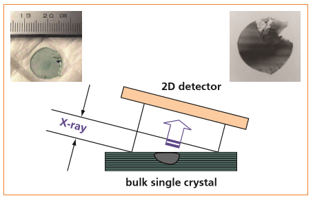
The XRT setup uses a standard sealed Cu X-ray tube. The specimen is irradiated with a beam generated by an X-ray mirror. Its axial divergence was limited. The PIXcel3D in 2D mode was applied to record the topographs.
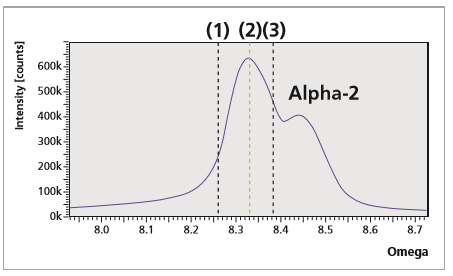
Topographs were taken on asymmetric grazing-incidence reflections of bulk single crystals at different angular positions of the rocking curve as indicated in the graph below. For recording rocking curves the PIXcel3D is simply switched to a receiving slit mode.
It then acts like a normal point detector with an electronically controlled active area.
The depicted example is from a c-axis oriented LiTaO3 crystal. The pictures on the right demonstrate the achievable image quality and resolution. The colors red to yellow indicate areas of high diffraction intensity. From the topographs the origin of the broadening of the widths of the rocking curves could be correlated to the curvature of the crystal surface.
(0 2 10) rocking curve of LiTaO3. Topographs (shown below) were taken at the indicated omega positions. The integration time was only 1 second (!) for each picture.

X-ray topography (XRT) was successfully demonstrated using the unique 2D performance of the PIXcel3D. High quality topographs with good resolution could be recorded from single crystals. The necessary integration time is typically in the order of seconds.
An XRT setup can be easily realized on X’Pert3 MRD and Empyrean instruments with commercially available components.