There is a requirement in many industries to identify and analyze crystals suspended in some sort of a medium using the technique of light microscopy. For example in the pharmaceutical industry, crystals of an active material (the drug) may be suspended in a carrier such as a cream for direct application to the skin. It is often necessary to characterize these suspended crystals for research and development purposes or from a quality control prospective. However, depending on the optical characteristics of the carrier, identifying the crystals may not be easily achieved by light microscopy alone due to lack of contrast between crystal and carrier.
Since crystals are birefingent and typically the carrier medium is not, the use of cross polarized microscopy often allows the crystals of interest to be identified. Typically analyses are performed manually, where an operator identifies individual crystals, and measures their size. This procedure can be time consuming, subjective depending on the operator's skill level and generally is not statistically significant.
This application note describes performing automated analyses using the Morphologi G3 fitted with crossed polarizer accessories, thereby addressing the issues of the manual method.
In this example, a pharmaceutical cream contains crystals of an active material. There is a requirement to identify and measure the length of crystals and then classify them according to their length.
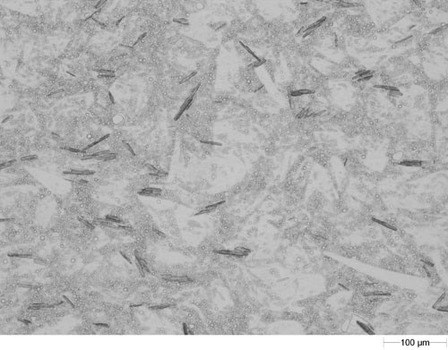
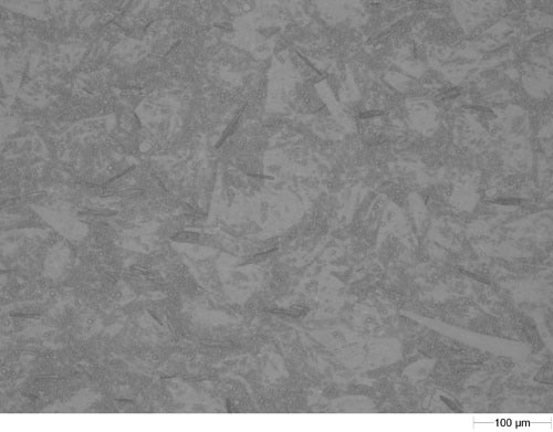
A small aliquot of the cream was sandwiched between two microscope slides to disperse it for presentation to the Morphologi G3. Images of the dispersed cream using both diascopic and episcopic light sources on the Morphologi G3 are shown in figure 1. When viewing the cream using light microcopy coupled with automated image analysis it is not simple to segment the crystals from the cream background since there is not sufficient contrast.
|
|
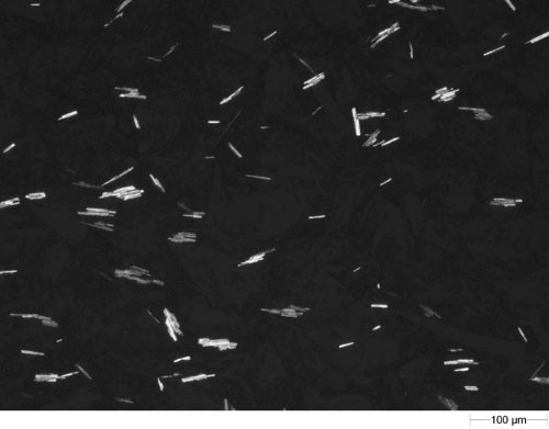
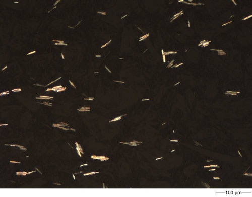
However, when crossed polarizers are placed in the light path, only the birefringent crystals can be seen as shown in figure 2 (both in gray scale and in color). Therefore there is now much better contrast between the crystal particles and the carrier which appears as a dark background and so the crystal particle images can now be separated from the background by image analysis.
|
|
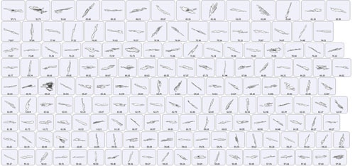
The Morphologi G3 software offers the option to analyze the particles automatically in grey scale or manually in color using the manual microscope with the grab and analyze feature. In this measurement a 4.9 x 10.8 mm scan area was analyzed automatically in around 20 minutes and a post analysis filter (solidity <0.7) was applied to exclude images of touching particles, resulting in almost 9000 particle being reported. Figure 3 shows examples images of particles after the automated analysis. The length of each particle is also displayed (microns). The user can return to particles of interest to view them in color.
|
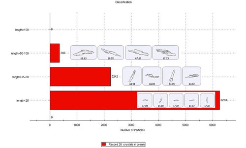
The results can be classified in terms of their size or other morphological parameters. For this application the particle images were classified according to their length in to the following classes:
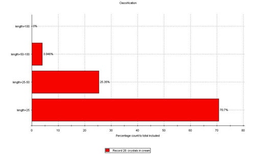
Figure 4 shows the result of the classification on a count basis, along with some example particle images. Figure 5 shows the same result in terms of a number based percentage of particles in each class.
|
|
The addition of polarizer accessories to the Morphologi G3 allows the automated identification and characterization of crystals suspended in a cream or other medium.
Such automated analysis reduces the operator's time and removes the subjectivity that can be associated with manual measurements. Additionally statistically significant numbers of particles can be measure in a short amount of time improving the reliability and reproducibility of results.