The Hydro SV allows measurement of a small volume (6-7ml) of dispersed sample. This is useful for cases when the supply of sample or dispersant is either limited or costly. In this application note we discuss the measurement of small sample volumes and, in particular, how repeatable, reproducible measurements can be obtained using the Hydro SV unit.
It can often be the case that the volume of sample available for particle size analysis is minimal. For example, in the pharmaceutical and catalysis industries it may be necessary to determine the particle size distribution early in the product development cycle, when only a few milligrams of material have been synthesized. This requires the ability to perform reliable measurements on a small volume of sample, and it is desirable to be able to recover the sample for further testing.
The principle challenge associated with performing laser diffraction measurements on small sample volumes is obtaining reproducible results. Both the sampling of the material and dispersion must be controlled in order to obtain meaningful data. This can be difficult to achieve when there are only a few milligrams of powder available for method development and analysis.
|
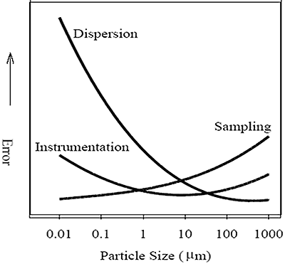
Sampling is controlled by the user selecting a representative sample for analysis and is the most significant source of error when measuring large particles (Figure 1). To obtain a representative result a minimum number of particles need to be measured. For example, to achieve a standard error of 5% on the Dx (90) requires at least 400 particles to be measured. Effectively this places an upper size limit, approximately 200µm, on the particle size that can be measured using small volume sample dispersion units, such as the Hydro SV.
Dispersion is controlled through the selection of an appropriate dispersant, stir speed and possibly through the application of external sonication. Dispersion is the greatest source of error when measuring fine particles (Figure 1).
The Hydro SV is a wet dispersion unit comprising a 6-7ml cuvette with magnetic stirrer. This allows the sample to be circulated through the measurement zone continuously, allowing multiple measurements to be carried out. The suspension of the sample can be observed directly in the cuvette or deduced from trends in the data. The contents of the cuvette can be decanted at the end of the experiment and the sample recovered.
To illustrate fully the capabilities of the Hydro SV it is necessary to test both the dispersion of a fine sample and the sampling of a relatively coarse material. These qualities are best shown by two different materials. A micronized lactose sample was measured to assess the dispersion process. This material is fine and therefore a challenge to disperse. A pigment sample containing larger particles was measured to illustrate that, with good sampling, reproducible results can be obtained in a small volume dispersion unit.
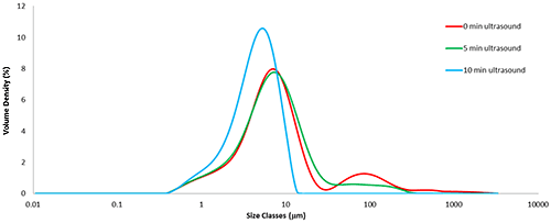
The micronized lactose sample was measured as received and following periods of sonication. To minimize the volume of the Hydro SV cuvette there is no internal ultrasound so it is applied externally, in this case using a sonic bath. The particle size distribution of the micronized lactose as received shows that the sample is agglomerated (Figure 2). After the first period of ultrasound, the particle size has decreased but there are still agglomerates present. Further sonication was therefore applied until the all of the agglomerates have been dispersed and the results have become stable.
|
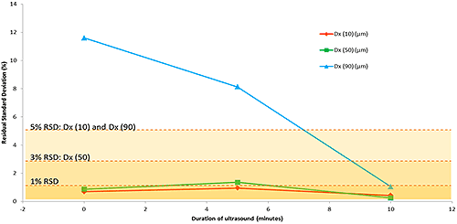
The particle size distributions shown in Figure 2 are average distributions created from five repeat measurements at each point in the dispersion process. Figure 3 shows the decrease in relative standard deviation (over the five repeat measurements) as the duration of ultrasound is increased. The higher variability before ultrasound, and after five minutes of ultrasound, indicate that the dispersion is not yet stable. After ten minutes of ultrasound the variability is found to be well-within the acceptable range, according to the ISO standard [2]. The reduced variability combined with the narrowed particle size distribution shows the sample has been fully dispersed.
|
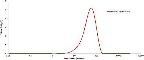
A green pigment sample containing larger particles was measured to illustrate that, with good sampling, reproducible results can be obtained in a small volume dispersion unit. A typical particle size distribution from the green pigment is shown in Figure 4. This sample is relatively large to be measured in the Hydro SV, with a Dx (90) of almost 100µm. For material of this size, sampling is the largest potential source of error (Figure 1).
|
For samples containing larger particles it is important to select a stir speed for the Hydro SV which is sufficient to ensure that the sample remains suspended and a representative sample of the material passes through the measurement zone in the cuvette. A stir speed of 1500-1800rpm is recommended for most powder samples.
The size distribution statistics obtained from six measurements of one aliquot of pigment shows the repeatability (Table 1A) of measurements to be well-within the guidelines of the ISO standard for laser diffraction [2]. The size distribution statistics obtained from six aliquots of pigment shows the reproducibility of the measurements. Reproducibility will be strongly influenced by the quality of the chosen sampling method. The data show that, with good sampling, the reproducibility achieved using the Hydro SV (Table 1B) also falls within ISO limits.
| A | Dx (10) (µm)
ISO 5% | Dx (50) (µm)
ISO 3% | Dx (90) (µm)
ISO 5% |
|---|---|---|---|
| A1-1 | 19.6 | 50.7 | 98.8 |
| A1-2 | 19.4 | 50.3 | 97.4 |
| A1-3 | 19.3 | 50.2 | 97.6 |
| A1-4 | 19.2 | 49.8 | 96.4 |
| A1-5 | 19.2 | 49.8 | 96.7 |
| A1-6 | 19.1 | 49.7 | 96.2 |
| Mean
1xStd Dev 1RSD (%) | 19.3
0.20 1.03 | 50.1
0.41 0.82 | 97.2
0.96 0.99 |
| B | Dx (10) (µm)
ISO 5% | Dx (50) (µm)
ISO 3% | Dx (90) (µm)
ISO 5% |
|---|---|---|---|
| Average of 'A1' | 19.1 | 49.7 | 96.6 |
| Average of 'A2' | 19.0 | 48.7 | 95.2 |
| Average of 'A3' | 21.3 | 52.2 | 99.6 |
| Average of 'A4' | 19.6 | 48.4 | 94.4 |
| Average of 'A5' | 19.8 | 50.5 | 100.0 |
| Average of 'A6' | 20.3 | 50.4 | 97.1 |
| Mean
1xStd Dev 1RSD (%) | 19.9
0.86 4.31 | 50.0
1.37 2.73 | 97.1
2.28 2.34 |
The ability to carry out routine measurements using small sample volumes is important in a variety of applications, especially when measuring chemical and pharmaceutical samples during early product development. In these cases, the Hydro SV can provide a robust means for making repeatable, reproducible measurements of very small volumes of sample.