Transmission experiments were performed on a hen egg-white lysozyme sample. The high angular resolution of the data enabled cell searching, indexing and unit cell refinement. The lattice parameters determined were in good agreement with the data obtained from single crystal measurements.
Diffraction experiments on protein samples have traditionally been relegated to single crystal diffraction systems for determining structure, or beam lines for extracting powder patterns from very small quantities of poorly diffracting material.
Diffraction experiments on protein samples have traditionally been relegated to single crystal diffraction systems for determining structure, or beam lines for extracting powder patterns from very small quantities of poorly diffracting material. The focusing mirror from Malvern Panalytical, in conjunction with solid-state line detectors like the X’Celerator or the PIXcel, has made it possible to perform these experiments in your own laboratory with Cu radiation. The size of the capillary tube no longer governs resolution with this optic, and peaks narrower than 0.05 degrees are easily resolved.
The ultra-fast solid-state line detectors make data collection from these poor diffractors possible in a reasonable amount of time, and give the ability to pre-screen and investigate macromolecular samples in your own laboratory using X-ray powder diffraction.
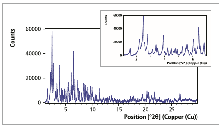
Background corrected scan of the tetragonal lysozyme crystals (16 hours measurement)
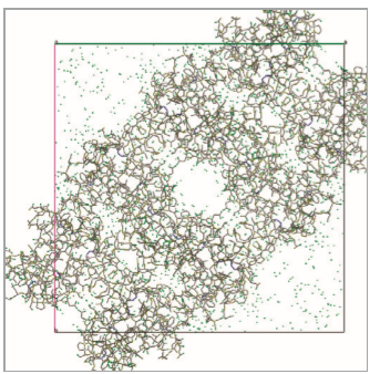
Protein crystals were grown at room temperature, from a solution consisting of protein, sodium acetate-acetic acid buffer and sodium chloride, at pH=4.8. The crystals in solution were then pipetted into a 0.5 mm capillary tube, and manually compacted.
This focusing mirror was used in conjunction with the PIXcel3D detector in scanning mode. The capillary spinner stage was used to spin the sample and randomize the protein crystal orientations. 2 hours scans were collected repeatedly over the course of 16 hours.
The experimental data were subject to crystallographic analysis. The resulting unit cell was compared with single crystal data.
Empyrean goniometer with focusing mirror, capillary spinner and PIXcel3D detector.
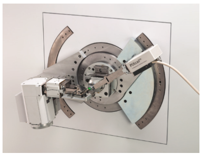
Empyrean capillary sample stage and Beam path of the used configuration

A cell search using HighScore Plus software yielded the following results using the DICVOL algorithm:
Tetragonal
a = 79.03 Å
c = 37.96 Å
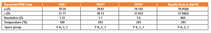
Further processing using a Pawley fitting method confirmed the unit-cell. Space group determination was done using the program ExtSym as implemented in HighScore Plus. The cell parameters and space group determined using the HighScore suite are in excellent agreement with the results from single crystal diffraction data.
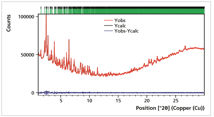
Pawley fit and space group determination of the lysozyme using the ExtSym program
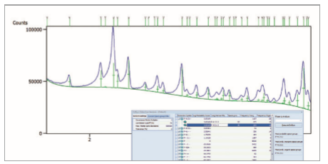
Pawley fitting of the lysozyme data using a Finger-Cox-Jephcoat asymmetry function. Cell parameters: a= b =78.95(8)Å and c = 37.93(4)Å and space group P43212.