
Fluorescence is commonly a problem in mineralogical phases like Hematite- Eskolaite, because these minerals contain the strongly fluorescing Cr and Fe ions. This data sheet shows how the 1Der detector excludes both Cr and Fe fluorescence. Empyrean’s unique optical configuration provides truly clean data leading to an excellent Rietveld refinement and resulting in an accurate crystal structure determination of this natural mineral.
For X-ray diffraction (XRD) measurements on the Empyrean system, it is beneficial to be able to choose an X-ray tube to suit the requirements of a material. Standard Empyrean tubes offer a choice of Cr, Mn, Fe, Co, Cu, Mo and Ag, and other anode emission energies can be provided on request. Cu is the most commonly used anode and the Cu Kα emission peak is the most commonly used energy for diffraction experiments.
In an X-ray diffraction experiment, the optics and the detector are together tuned to the narrow energy window required by the experiment. The 1Der strip detector can be automatically tuned to provide a narrow energy window around any chosen diffraction energy. This provides full flexibility across the whole range of Empyrean 1D applications.
All elements fluoresce when irradiated with a certain X-ray energy. In a diffraction measurement, fluorescence from some elements can give rise to unwanted background intensity in a diffraction pattern. A high background reduces the possibility of identifying the low intensity peaks that often provide the final clues in a Rietveld crystal structure refinement. Figure 1 illustrates the elements that emit the greatest fluorescence interference in a diffraction measurement using a Cu X-ray tube. There are various strategies for excluding fluorescence interference and, in this datasheet, we show how the 1Der detector address the double challenge of fluorescence from both Cr and Fe elements.
For X-ray diffraction (XRD) measurements, it is beneficial to be able to choose an X-ray tube to suit the requirements of a material. Standard Empyrean tubes offer a choice of Cr, Mn, Fe, Co, Cu, Mo or Ag. Other anode emission energies can be provided on request. Cu is the most commonly used anode and the Cu Kα emission peak is the most commonly used energy for diffraction experiments.
Anode choice depends upon the requirements of the measurement, for example, spreading a diffraction pattern using low energy (long wavelength) emission (e.g. Cr, Mn), provides improved peak separations for organic materials; condensing a pattern, using a high energy (short wavelength) emission (Mo, or Ag) provides full total scattering data for PDF analysis and high sample penetration.
In an X-ray diffraction experiment, the optics and the detector are together tuned to the narrow energy window required by the experiment. The 1Der strip detector can be automatically tuned to provide a narrow energy window around any chosen diffraction energy. This provides full flexibility across the whole range of Empyrean 1D applications.
All elements fluoresce when irradiated with a certain X-ray energy. In a diffraction measurement, fluorescence from some elements can give rise to unwanted background intensity in a diffraction pattern. A high background reduces the possibility of identifying the low intensity peaks that often provide the final clues in a Rietveld crystal structure refinement. Figure 1 illustrates the elements that emit the greatest fluorescence interference in a diffraction measurement using a Cu X-ray tube. There are various strategies for excluding fluorescence interference and, in this datasheet, we show how the 1Der detector address the double challenge of fluorescence from both Cr and Fe elements.
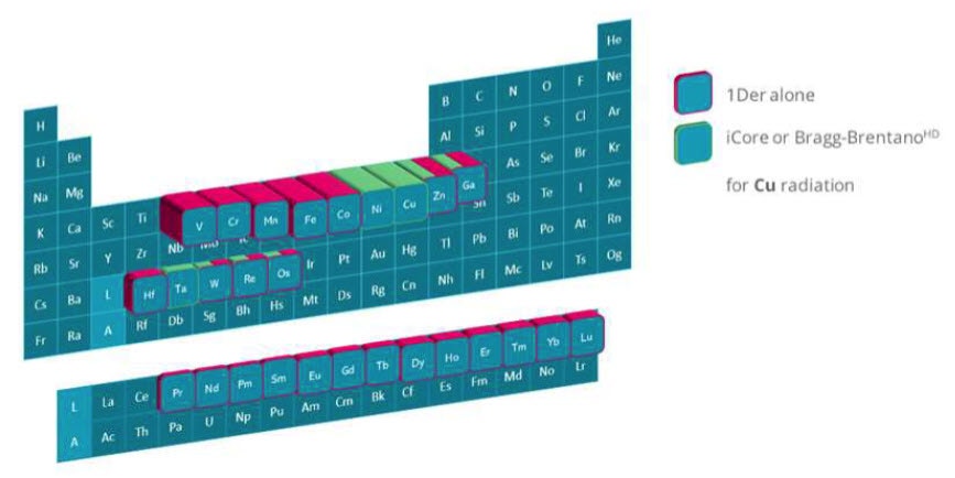
Figure 1. The periodic table of elements showing elements that are easily excited by X-rays from a Cu-X-ray tube and the optical configurations that can be used to exclude their fluorescence are indicated. Alone, the 1Der excludes unwanted fluorescence from Cr and Fe. An equivalent picture can be obtained for each X-ray source wavelength. For example, with a Co X-ray source the row of fluorescing elements will extend from Sc to Cu.
Hematite and Eskolaite are naturally occurring minerals and endmembers of a solid solution that exhibits complex magnetic phase diagram as function of temperature.
Crystal structure refinement using the Rietveld method is used to gain insight into the phase stability of similar minerals and comparable synthetic materials. This solid solution is also interesting as a model system for testing the capabilities of XRD systems to exclude fluorescence interference from data. A powder sample with a mixed Hematite-Eskolaite composition, namely (1-x)Cr2O3-xFe2O3 with x = 0.3 provides a fluorescence challenge because both Cr and Fe are fluorescing elements.
The Co Kα emission energy is insufficient to excite fluorescence from Fe-containing samples, so Co X-ray tubes are often used for Fe-containing samples to obtain clean data with a low background. However, Co radiation is energetic enough to cause strong Cr fluorescence.
The data shown in Figure 2 were obtained with a Cu X-ray tube, iCore incident beam optics (in fixed slit mode), dCore diffracted beam optics (with fixed anti-scatter slit) and the 1Der detector. The sample was presented in the reflection-transmission spinner stage and measured in Bragg-Brentano reflection geometry. As shown in Figure 1, the 1Der detector with its narrow energy window, totally excludes fluorescence from both elements. This enabled measurement of clean data (as shown by the red line in Figure 2) with a low background sufficient to achieve excellent Rietveld refinement including the weakest diffraction peaks (see inset).
The data were analyzed using HighScore Plus software [1]. Figure 2 shows the results of Rietveld refinement (blue and black lines) providing in an accurate crystal structure determination.
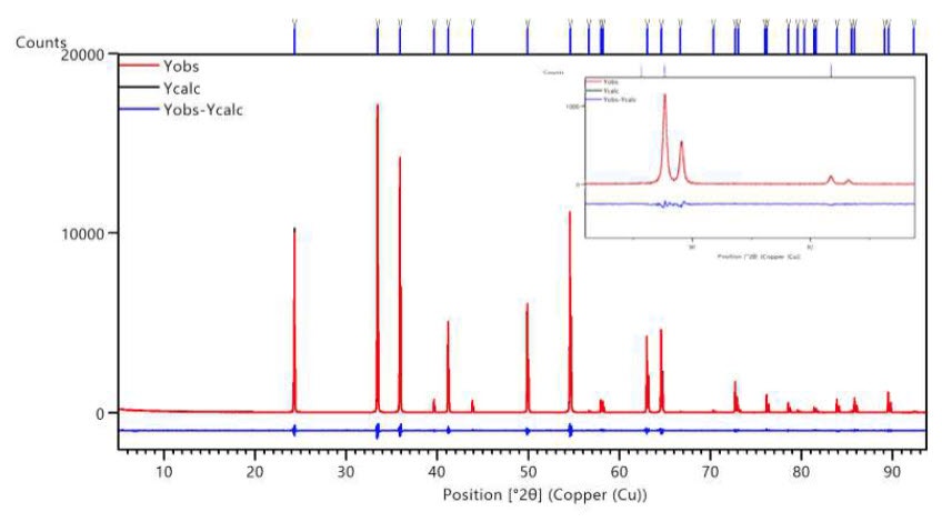
Figure 2. Rietveld refinement of (1-x)Cr2O3-xFe2O3 with x = 0.3; GoF = 1.07, Rwp =9.9%. The inset further demonstrates the excellent fit and very low noise level even at high 2θ. The calculated line in black is almost fully hidden underneath the measured data in red showing a very good refinement quality. The difference between the two lines is shown underneath in blue. The modelled peaks are indicated by blue vertical tags.
Using the unique Empyrean incident beam optics together with the 1Der detector, excellent data quality is achieved for XRD measurements of fluorescing materials. The data cleanliness is such as to include the weak but significant signal from low intensity peaks and to enable an all-time low Rietveld fit value for this material. 1Der has opened the possibilities further on an already competent platform. With 1Der you can truly exclude fluorescence interference and discover more about your materials.
[1] The HighScore Suite, T. Degen, M. Sadki, E. Bron, U. König and G. Nénert, Powder Diffraction 29 (S2), December 2014, S13-S18
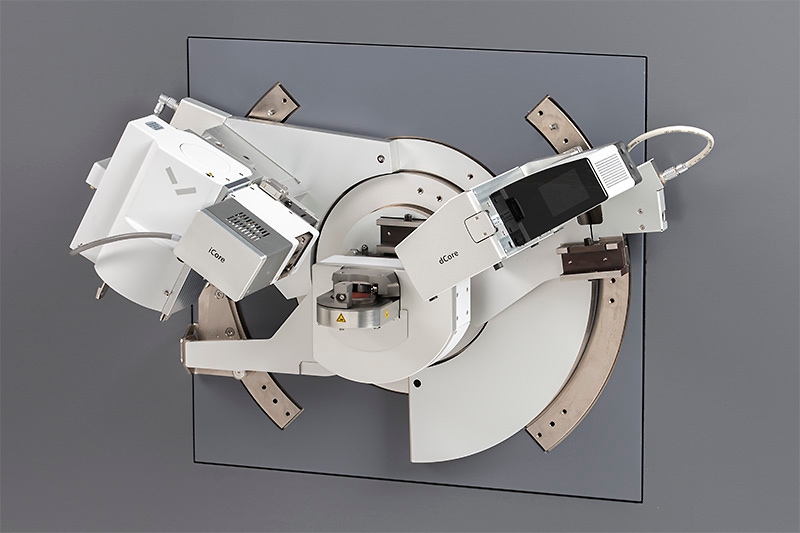
iCore + dCore configuration with the 1Der detector.