X-ray diffraction (XRD) is a powerful tool for studying cultural heritage objects. By identifying crystalline pigments, it can determine the materials used, whether alteration or corrosion has taken place, how the objects were made, and their originality. To protect and preserve the artifacts, this analysis must be non-destructive.
When working with smaller-sized objects, researchers often need to analyze on a sub-millimeter scale. Microdiffraction is ideal for these cases. This application note outlines the process and results of non-destructive microdiffraction with the Empyrean diffractometer on the pigments in a parchment leaf from a medieval Book of Hours.
Books of Hours were Christian devotional books used by wealthy men and women, typically containing a calendar of church feasts, excerpts from the Bible, psalms, and other prayers. They were produced in France, Belgium, the southern Netherlands, southern and western Germany, northern Spain, and southern England. Paris and its surroundings were a major center of production.
Known examples of these parchments date from the 13th to the early 16th century (when they were replaced by printed books) and range from simple to elaborate. The parchment leaf tested was written and painted around 1470 in Paris, France, and contains the ‘Obsecro te’, a 14-15th century prayer to the Virgin Maria. It is richly decorated, from a private collection, and measures 163 x 123 mm.
Login to access the full application note
X-ray diffraction (XRD) is a powerful tool for studying cultural heritage objects. By identifying crystalline pigments, it can determine the materials used, whether alteration or corrosion has taken place, how the objects were made, and their originality. To protect and preserve the artifacts, this analysis must be non-destructive.
When working with smaller-sized objects, researchers often need to analyze on a sub-millimeter scale. Microdiffraction is ideal for these cases. This application note outlines the process and results of non-destructive microdiffraction with the Empyrean diffractometer on the pigments in a parchment leaf from a medieval Book of Hours.
Books of Hours were Christian devotional books used by wealthy men and women, typically containing a calendar of church feasts, excerpts from the Bible, psalms, and other prayers. They were produced in France, Belgium, the southern Netherlands, southern and western Germany, northern Spain, and southern England. Paris and its surroundings were a major center of production.
Known examples of these parchments date from the 13th to the early 16th century (when they were replaced by printed books) and range from simple to elaborate. The parchment leaf tested was written and painted around 1470 in Paris, France, and contains the ‘Obsecro te’, a 14-15th century prayer to the Virgin Maria. It is richly decorated, from a private collection, and measures 163 x 123 mm (Figure 1.)
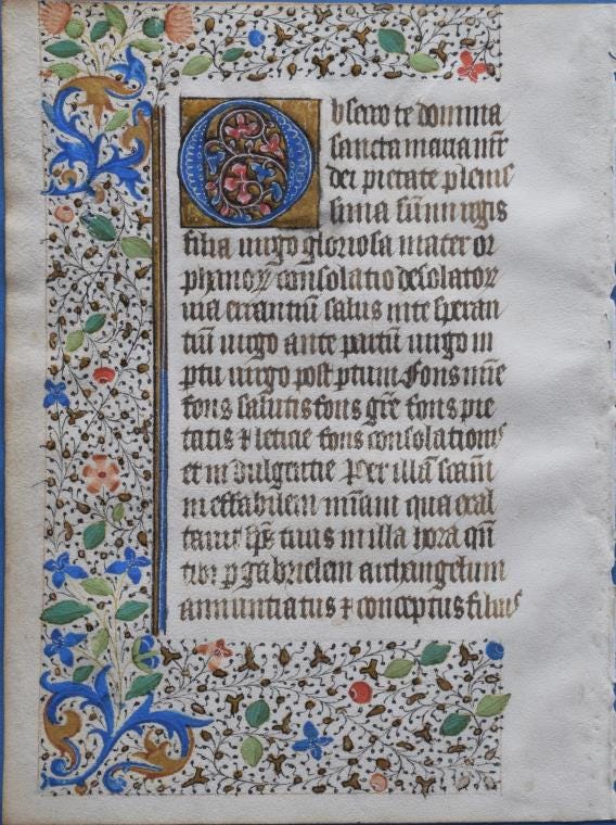
Figure 1: The decorated parchment
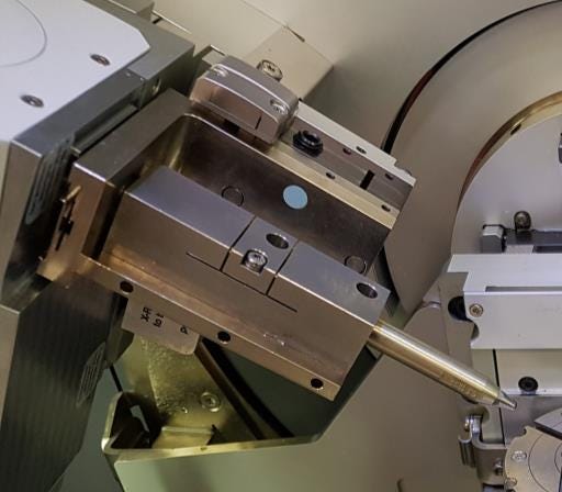
The parchment’s pigments were analyzed using microdiffraction on the Empyrean diffractometer, a multipurpose system equipped with (Figure 2):
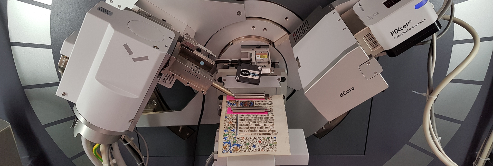
Figure 2: The experimental setup in the Empyrean
The parchment leaf contained fine detail: for instance, a white line within the large initial was only 0.3 mm wide. To ensure a spot width of 0.3 mm, a 135-millimeter-long monocapillary with a 0.1-millimeter opening was used (Figure 3 and 4). X-rays were transported through the monocapillary using the effect of total reflection inside the capillary, resulting in a narrow X-ray beam. Spots of each pigment color were analyzed in this way, with a measurement time per spot of 18 minutes.
|
Figure 3: The monocapillary with component recognition, mounted with the PreFIX system in the Empyrean diffractometer.
|
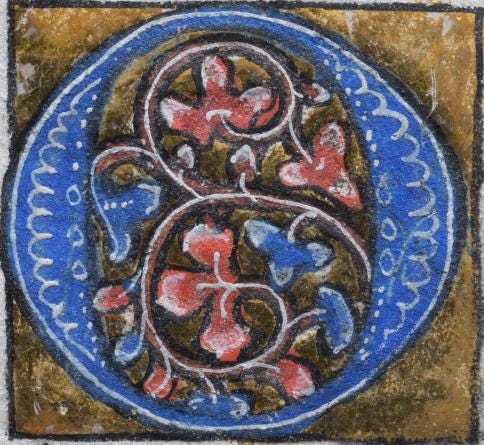
Figure 4: The large initial, 26 x 27 mm, decorated with blue and red colors and gold leaf. The white lines are 0.3 mm wide.
|
Blue pigment:
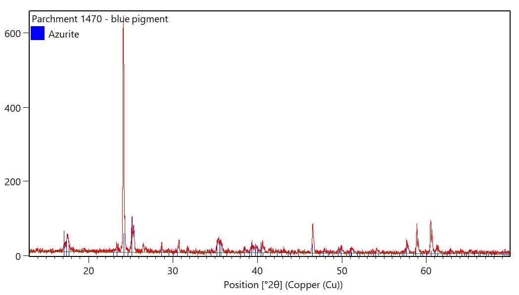
The analysis identified the blue pigment as azurite, Cu3(CO3)2(OH)2 (Figure 5 and 6). Azurite is a natural pigment and was available in the medieval period. It can be found on several localities in France and other countries. Azurite in a good pigment quality is expensive and must be prepared in the right grain size, becoming bluish-grey if grains are too fine. Other blue pigments used in medieval times are lazurite, well known since antiquity as the blue component in the gem material lapis lazuli, and indigo, obtained from plants.
|
Figure 5: Microdiffraction measurement of a spot on the blue pigment, identified as azurite.
|
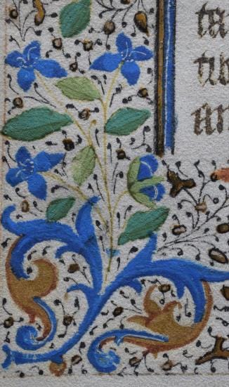
Figure 6: Detail from the parchment with blue and green pigments (55 x 34 mm).
|
Green pigment:
The dark green pigment was identified as malachite, Cu
2
(CO
3
)(OH)
2
, and an unknown compound. Malachite was available in the medieval period. It can be natural or artificial: the poor crystallinity according to the measured data may indicate a synthetic pigment. Other green pigments used in the medieval period are copper acetate (artificial), copper chloride (artificial), copper hydroxy-sulfates (artificial), or earthy iron silicates (natural).
Red pigment:
The red pigment was identified as cinnabar, HgS (Figure 7). This is a natural pigment found in Almaden, Spain, and other locations, and was well known in the medieval period. The low amount of quartz found also indicates a natural material. Another red pigment commonly used in these times is minimum, Pb
3
O
4
.
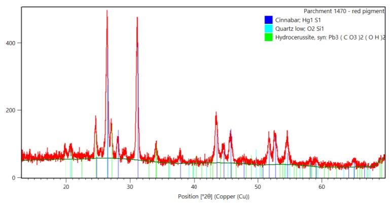
Figure 7: Microdiffraction measurement of a spot on the red pigment: cinnabar with some quartz and hydrocerussite.
White pigment:
The analysis identified the white pigment as hydrocerussite, Pb
3
(CO
3
)
2
(OH)
2
, with an admixture of cerussite, PbCO
3
(Figure 8). This is a synthetic pigment known as ‘white lead’. It is produced from metallic lead under the influence of vapor of acetic acid in a CO
2
-rich environment. The procedure for the preparation of white lead pigment was known since antiquity. It is a very common pigment in medieval times.
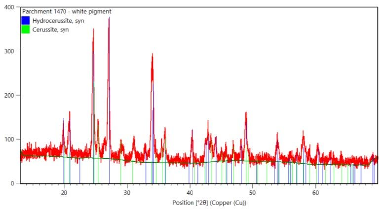
Figure 8: Microdiffraction measurement of a spot on the white pigment, identified as hydrocerussite with cerussite, known as ‘white lead’.
Gold leaf:
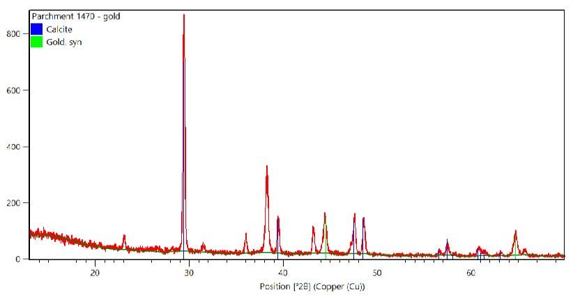
The analysis identified the gold leaf spot as gold, Au, with calcite, CaCO
3
(Figure 9 and 10).
|
Figure 9: Microdiffraction measurement of a spot on the gold leaf, showing gold and the underlying calcite (calcium carbonate, probably chalk) primer. Figure 10: Detail with gold leaf and calcite primer (11 x 6 mm).
|
Figure 10: Detail with gold leaf and calcite primer (11 x 6 mm)
|
Based on these results, we can draw conclusions about the parchment’s authenticity and manufacture. Firstly, all the pigments were identified as materials typically used in the medieval period. The analysis did not identify any materials that were unavailable in the medieval period, such as white titanium oxide, a white pigment not produced until 1916. Based on this analysis, there is no indication that the parchment leaf is a fake.
Secondly, the results also provide insight into the production of the parchment leaf. For example, the identification of calcite in the gold leaf spot suggests that a fine primer of calcium carbonate (probably chalk) was put on the parchment first to obtain a smooth, flat surface, and the gold leaf was placed on it afterward.
This application note demonstrates that the Empyrean diffractometer is an ideal multipurpose instrument for the analysis of smaller-sized cultural heritage objects. By supporting X-ray microdiffraction using monocapillaries or focusing lenses, it enables non-destructive analysis of fine details on a sub-millimeter scale. In turn, this can facilitate detailed archaeological research on a wider range of objects.
![[Figure 9 AN210714-Empyrean-Medieval-Parchment.png] Figure 9 AN210714-Empyrean-Medieval-Parchment.png](https://dam.malvernpanalytical.com/01c6e0f3-2cbc-467f-9623-aef2008c6959/Figure%209%20AN210714-Empyrean-Medieval-Parchment_Original%20file.png)