

Looking for more information?
To request a quote, more information or download a brochure select an option below.
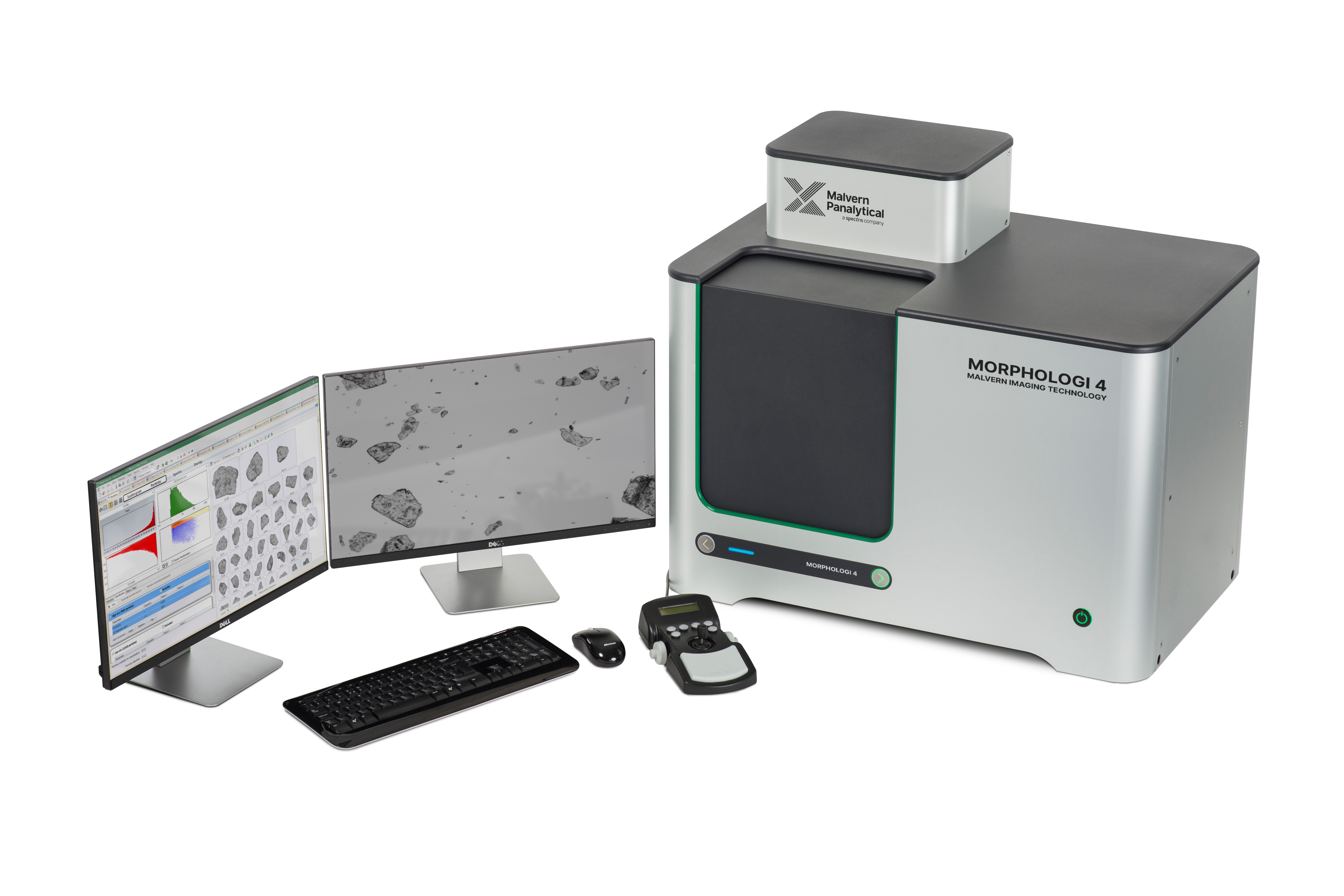
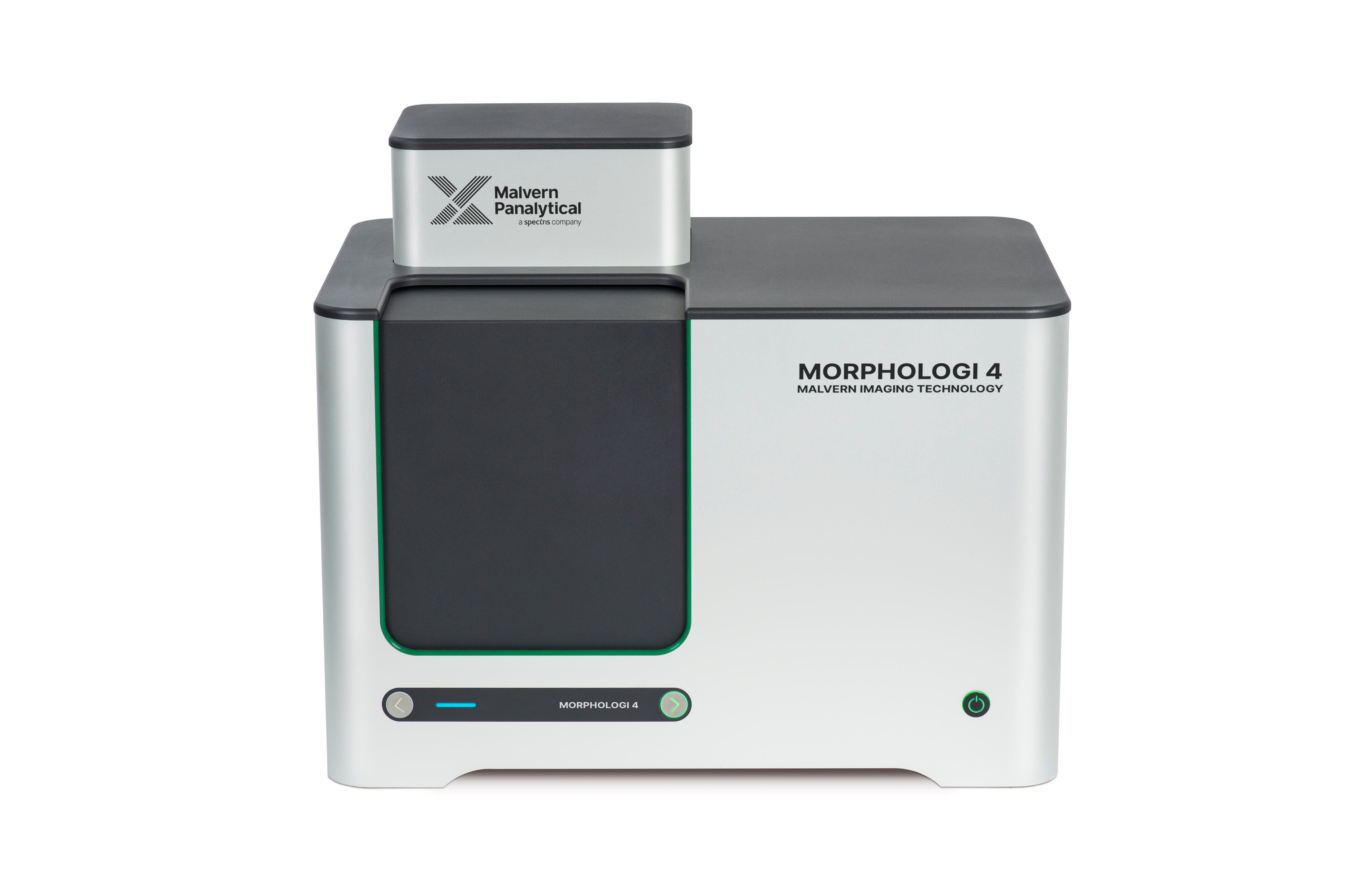
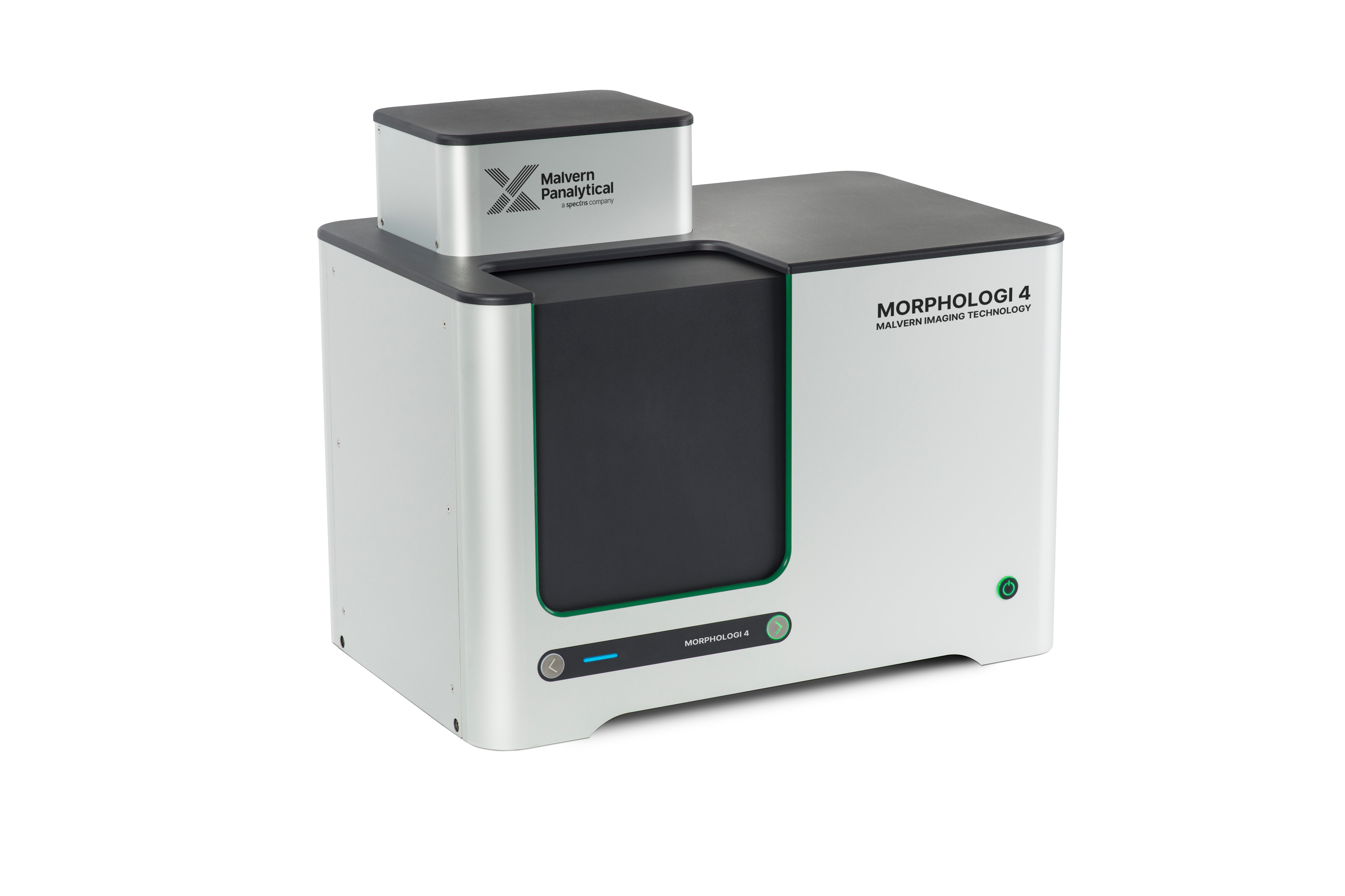
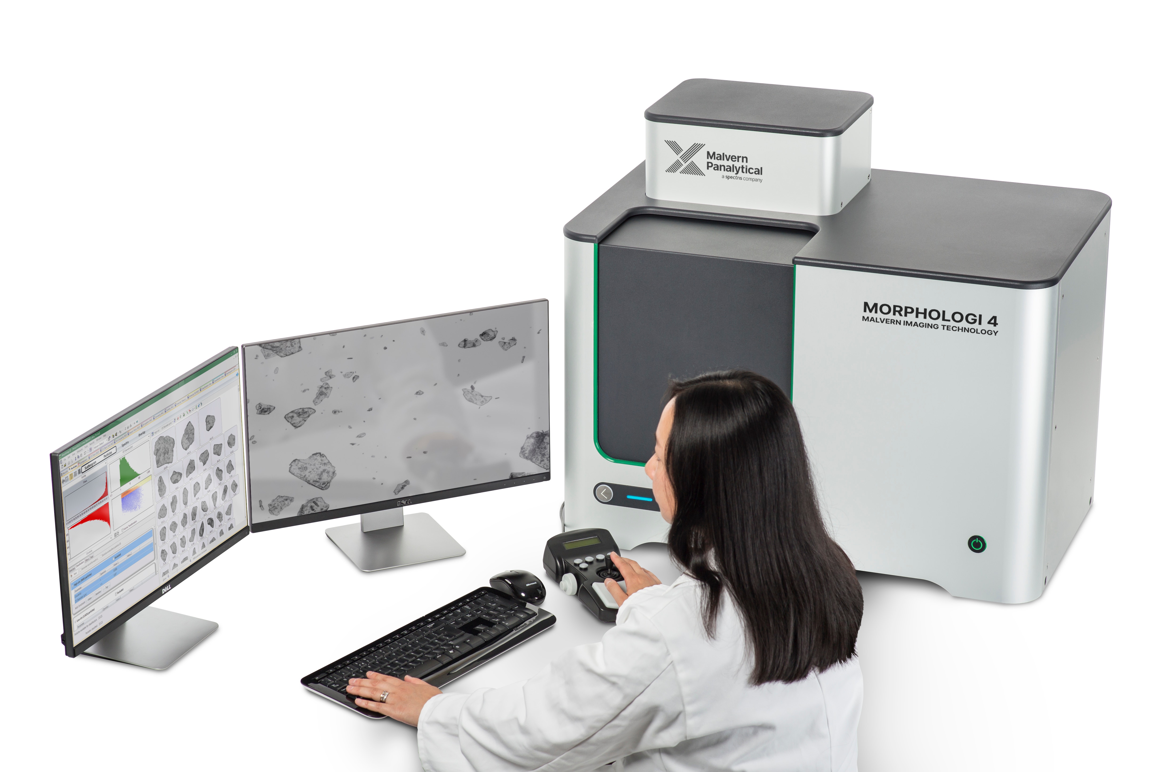
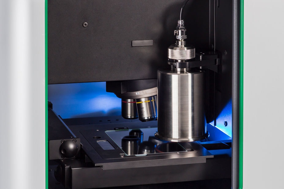
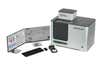
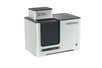
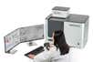
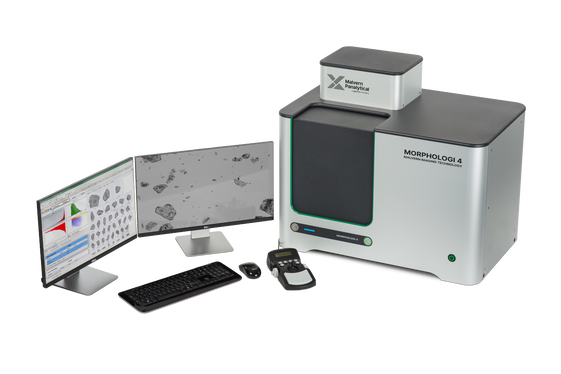
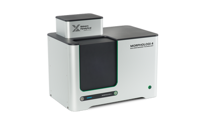
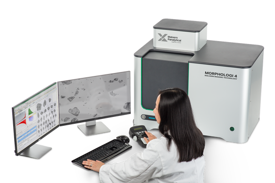
Morphologi 4 provides detailed morphological descriptions of particulate samples through static image analysis, enabling a greater understanding of both sample and process.
It can equally be used as an R&D tool to investigate challenging applications and in automated QC analysis where generation of robust, user-independent results and validation are required.
Broad particle size range, from 0.5 μm to over >1300 μm, enables size measurements of a wide range of samples
20+ morphological parameters deliver a highly-detailed description for deeper understanding of your particulate material
Advanced manual microscope mode and ability to return to particles of interest enables an even closer examination of unexpected particles
High-resolution microscope ensures quality particle images for optimum image analysis data
Integrated dry powder dispersion unit delivers reproducible sample dispersion, critical to achieving meaningful results
SOP control, from sample dispersion to data analysis, provides simple and automated operation for robust, repeatable measurements
Dedicated sample presentation accessories enable measurement of a wide variety of sample types, including suspensions and filters
The only instrument that can measure the particle size range from low to high for polymer particles dispersed in water.
Ron Peters — DSM
| Technology | Static automated imaging |
|---|---|
| Particle size | 0.5 μm – 1300 μm (upper limit may be extended for some applications*)
* Sample and substrate dependent |
| Particle properties measured | Size, shape, transparency, count, location |
| Particle size parameters | Circle equivalent (CE) diameter, length, width, perimeter, area, maximum distance, sphere equivalent (SE) volume, fiber total length, fiber width |
| Particle shape parameters | Aspect ratio, circularity, convexity, elongation, high sensitivity (HS) circularity, solidity, fiber elongation, fiber straightness |
| Particle transparency parameters | Intensity mean, intensity standard deviation |
| Integrated Sample Dispersion Unit | For fully automated dispersion and measurement of dry powders. Manual or SOP control of dispersion pressure, injection time and settling time |
| Illumination | White light LED: brightfield, diascopic and episcopic; darkfield, episcopic |
| Detector | 18 MP; 4912 x 3684 pixel color CMOS array; pixel size 1.25 μm x 1.25 μm |
| Optical system | Nikon CFI 60 brightfield / darkfield system |
| Lens (and particle size range) | 2.5x: 8.5 µm – 1300 µm (nominal)
5x: 4.5 µm – 520 µm (nominal)
10x: 2.5 µm – 260 µm (nominal)
20x: 1.5 µm – 130 µm (nominal)
50x: 0.5 µm – 50 µm (nominal) |
| Dimensions | 810 mm (W) x 520 mm (D) x 685 mm (H)
with carry handles: 1100mm (W) x 520mm (D) x 685mm (H) |
|---|---|
| Weight | 76 kg (84kg including carry handles) |
| Power | 100-240 V ac 50/60 Hz (<100W load) |
Appropriate dispersion of the individual particles and agglomerates within a sample leads to robust and reliable results. Good spatial separation and representative sampling of the particles is required. To achieve this, a number of dispersion options are available with the Morphologi 4.
Easy, reproducible, automated sample dispersion
Morphologi 4 comes with an integrated dry powder dispersion unit. It enables simple, reproducible preparation of dry powder samples. A precise amount of sample is dispersed automatically using a unique compressed air dispersion mechanism controlled from within the software, for reproducible dispersions every time.
Optional accessories – for analysis of particles in suspension
A range of instrument accessories supports preparation of samples on microscope slides, in wet suspensions or on filters, extending the capability of the Morphologi 4 beyond the characterization of dry sample dispersions. Each accessory fits directly into the automated stage area and is easily selectable in the Morphologi software.
The Morphologi 4 measurement procedure can be split into four sections:
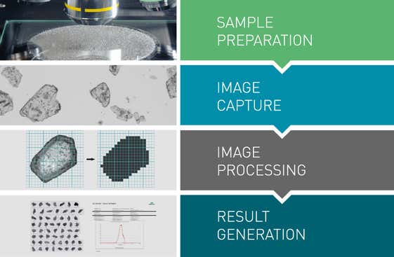
Spatial separation of individual particles and agglomerates is critical to representative results. The integrated dry powder disperser makes preparing dry powder samples easy and reproducible. The applied dispersion energy can be precisely controlled, enabling the measurement process to be optimized for a range of material types. Dispersion is achieved without explosively shocking the particles, avoiding the damage of fragile particles whilst ensuring strongly-agglomerated materials are dispersed. Accessories that fit directly in to the Morphologi 4’s automated stage are available for preparing suspended or filtered samples.
The instrument captures images of individual particles by scanning the sample underneath the microscope optics. The Morphologi 4 can illuminate the sample from below or above, whilst accurately controlling the light levels.
Use of either the automated ‘Sharp Edge’ segmentation analysis or the manually-controlled thresholding enables the detection of particles and the calculation of a range of morphological properties for each.
Statistically representative distributions are constructed for each parameter from the thousands of particles that have been analyzed. Advanced graphing and data classification options in the software ensure that extracting the relevant data from the measurement is straightforward, via an intuitive visual interface. Individually-stored grayscale images for each particle provide qualitative verification of the quantitative results.
The only instrument that can measure the particle size range from low to high for polymer particles dispersed in water.
Ron Peters — DSM
Morphologi 4 provides value in a wide range of applications, some of which are described below:
Pharmaceutical development
Active Pharmaceutical Ingredient (API) and excipient particle size and shape are critical material attributes which must be understood and controlled during formulation development and process scale-up for both innovator and generic products. Morphologi 4 delivers the statistically-relevant particle size and shape information to enable this.
Spray drying
Spray drying techniques are employed across a range of manufacturing industries because of the high fluidity of the granules that they produce. This fluidity comes from the tight particle size distributions and specific particle shapes that are produced by the process. These morphological attributes can be monitored by Morphologi 4, which provides statistically-relevant data to support the optimization of the process and to enable manufacturers to confidently specify their product.
Energy storage/Batteries
Controlling the application of the electrode material in the battery manufacturing process, and the resulting film density and uniformity, is essential in optimizing battery power and energy storage . Morphologi 4 enables particle size and shape descriptions of the electrode material powder to be correlated with battery performance, to support product development and to ensure final product quality.
Powder metallurgy
Metal powder properties, such as particle size and shape, are critical for powder metallurgy processes as they impact final component quality and performance. Morphologi is used by metal powder producers and component manufacturers to ensure an optimum, consistent and traceable powder supply with the desired packing and flow characteristics, to mitigate the risk of costly part failure.
Mining and minerals
The morphology of geological deposits helps trace the extent of natural disasters and predict the impact of future environmental events. Particle properties also determine the effectiveness of abrasive minerals for use in cutting and polishing tools. Morphologi 4 automatically generates the statistically-relevant particle data required to meet these challenges.
The only instrument that can measure the particle size range from low to high for polymer particles dispersed in water.
Ron Peters — DSM
| Title | Version | Date | Download |
|---|---|---|---|
| {{ row.title }} | {{ row.softwareVersion }} | {{ row.date }} | Download |
Sorry, no manuals are available for this product
| Title | Version | Date | Download |
|---|---|---|---|
| {{ row.title }} | {{ row.softwareVersion }} | {{ row.date }} | Download |
Sorry, no software downloads are available for this product
To assure that your instrument remains in top condition and performs on the highest level, Malvern Panalytical offers a wide range of services. Our expertise and support services assure an optimal functioning of your instrument.
Service for a lifetime
Adding value to your processes

Static image analysis ideal for R&D and QC. Automate your manual microscopy with advanced software and particle analysis with accessories for every sample.


