A study by the Academic Laboratory of Materials Research of Paintings (ALMA) in Prague, Czech Republic has shown that X-ray powder micro-diffraction can be used for the investigation of salt damage. The use of X-ray microdiffraction enabled the research team to identify the probable causes of damage to an ancient fresco.
Pigment degradation, often resulting in color changes, causes serious damage to wall paintings. Many factors lead to pigment degradation including sunlight, air pollution, salts and microorganisms. Of these, salts are one of the most damaging agents, especially for artworks of a porous nature such as wall paintings.
Pigment degradation, often resulting in color changes, causes serious damage to wall paintings. Many factors lead to pigment degradation including sunlight, air pollution, salts and micro- organisms. Of these, salts are one of the most damaging agents, especially for artworks of a porous nature such as wall paintings.
The damage that salt causes to wall paintings is not only mechanical: it also causes chemical and mineralogical alterations to the painted surface. In order to select the optimum restoration procedure it is crucial to know what salt is present and its source.
A recent study by the Academic Laboratory of Materials Research of Paintings (ALMA) in Prague, Czech Republic has shown that X-ray powder micro-diffraction can be used for the investigation of salt damage. The use of X-ray micro- diffraction enabled the research team to identify the probable causes of damage to an ancient fresco.
The identification of pigments in artworks is essential if solutions to the problems of restoration, conservation, dating, provenance and authentication are to be found. X-ray micro-diffraction has emerged as one of the most appropriate techniques for such research, proving invaluable with its ability to distinguish inorganic pigments and reveal the products of degradation.
Given the historic importance of the St. George fresco, the use of non- destructive testing was essential. X-ray powder micro-diffraction allows samples to be archived after analysis or preserved for additional testing, if required. Furthermore, it requires only very small sample sizes, 1 mm or less, an important consideration when testing rare and valuable artwork.
Figure 1: Typical sample of a cross-section of a paint layer prepared in a perspex block
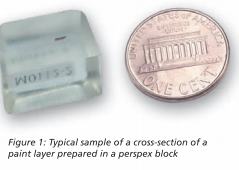
Samples for testing were removed from the wall painting by art restoration experts. The samples taken - fragments and cross sections - were just 1 mm in size (see Figure 1). A light microscope was used at a magnification of 50x for visual examination of the fresco.
The identification of single or multiple phases in an unknown sample is the main application of X-ray powder diffractometry. Phase identification is widely used in many fields, for example: identification of minerals in geological samples; detection of polymorphs in the pharmaceutical industry; determination of impurities in a pure phase; and forensic applications.
Malvern Panalytical solutions for phase identification include the Empyrean system, the X’Pert Powder and the CubiX . All are equipped with vertical goniometers. Malvern Panalytical's X’Pert HighScore software package is designed specifically for phase identification and enables the easy analysis of complex phase mixtures.
A Malvern Panalytical Theta-Theta (radius 240 mm) X-ray diffractometer was used to provide direct phase analysis of paint layers. The instrument was fitted with a cobalt X-ray tube and operated at 40 kV and 30 mA. The setup included a mono-capillary, with an exit diameter of 0.1 mm, providing a parallel X-ray beam and Malvern Panalytical's line detector X’Celerator.
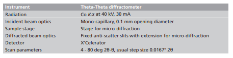
Figure 2 shows samples taken from the dark brown parts of the wall paintings (the scene boundary and figure clothing) viewed in an optical microscope under visible (white) light. The fragments consist of a dark brown layer on the surface with a white layer underneath.
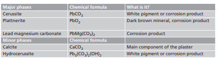
X-ray micro-diffraction results shown in Figure 3 and Table 1 confirm the presence of several different lead compounds: hydrocerussite, cerussite, plattnerite and lead magnesium carbonate. Both hydrocerussite and cerussite were widely used as a pigment called lead white. Plattnerite which causes the dark brown color of the painting, and lead magnesium carbonate were never used as pigments; they are corrosion products of the original pigment.
The formation of corrosion products can be associated with the degradation of two lead-based pigments – lead white and red lead (minium, Pb3O4).
1. Lead white - A potent oxidant is required to convert stable lead white into plattnerite. In the case of the wall paintings, oxidants must have arisen from an external source. For example some species of bacteria can oxidize hydrocerussite to plattnerite by means of hydrogen peroxide generation.
2. Minium - Although it has been known since antiquity that minium on wall paintings turns black with exposure to air, light and humidity, the mechanism of minium darkening is still not completely understood.
To test the minium theory, the ALMA team added magnesium cations and carbonates (ions that are very common in plasters) into a suspension of minium in water. After several weeks the color of the mixture has been darkened from orange to brown. The analysis of the product by X-ray powder diffraction showed that, as well as minium, plattnerite, cerussite and lead magnesium carbonate were present – the same compounds that are found in the real microsamples. Therefore the spontaneous transformation of minium into plattnerite and the presence of magnesium and carbonate ions are the most likely cause of damage to the St. George fresco.
Figure 2: Optical microscope photographs of samples from the dark brown parts of the wall paintings
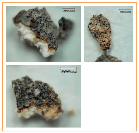
Figure 3: Micro-diffraction result of a wall paint sample
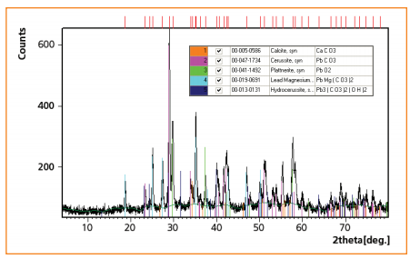
Table 1 Phases found in the sample
Samples were taken from two different areas of the fresco for the investigation of the origin and composition of salt efflorescence.
Diffractograms of the fragment surface and two spots on the cross section: Calcite and vaterite were identified in fragment and cross sectional samples taken from the same area of the wall painting. Vaterite is a metastable polymorph of calcium carbonate. It is rarely found in nature but can be formed by bacterial activity. Gypsum, a product of salt efflorescence, was found on the upper side of the fragment only.
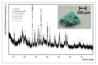
Diffractogram of blue-green fragment of wall painting: This sample contained: calcite – the main component of plaster under the painting layer; the pigments brochantite, malachite and hydrocerussite; and syngenite. Syngenite is a less common salt usually formed by the recrystallization of sulphates under the influence of potassium dissolved in ainwater.
Figure 4: Diffractograms of upper side of fragment and two spots on the cross-section
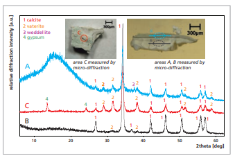
Figure 5: Diffractogram of blue- green fragment of wall painting
X-ray powder micro-diffraction is a non-destructive method of direct phase analysis. This application note demonstrates that the technique is ideal for the investigation of secondary crystalline phases in wall painting samples. X-ray micro-diffraction is the only method capable of producing such exact descriptions of the diverse phases with the same or similar elemental composition present in the fresco. This information improves understanding of the processes of pigment degradation, invaluable for artwork restoration and conservation.
The results of this study indicate that damage to the St. George fresco was probably caused by (i) oxidation arisen from hydrogen peroxide generated by bacteria, (ii) spontaneous transformation of minium into plattnerite and (iii) reaction of the pigments with magnesium cations and carbonates, commonly present in the plaster. Efflorescences containing gypsum and syngenite indicate the influence of rainwater on the wall painting, while vaterite is a sign of biological activity.