Advanced Particle Characterization

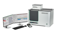
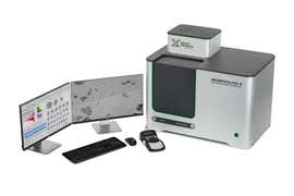
Image analyzers characterize particles in terms of their size, shape and transparency. They are usually categorized as either dynamic or static imaging systems. In dynamic image analysis systems, the particles flow past the imaging optics, whereas in static image analysis systems, the sample being measured is dispersed and remains stationary for presentation to the imaging system. Malvern Panalytical’s Morphologi Range comprises automated static image analyzers, which provide excellent, non-subjective, hands-free analysis. The ‘ID’ variant of the Morphologi system includes a Raman spectrometer, which provides component-specific chemical identification in addition to particle size and particle shape, using the technique of Morphologically-Directed Raman Spectroscopy (MDRS®).


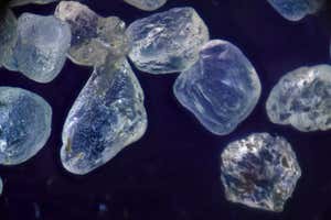
Image analyzers provide statistically-relevant data on particle shape and size for hundreds or even thousands of particles per measurement. This enables deeper understanding of the whole sample and allows you to determine if your sample contains single particles or agglomerates, whether your particles are regular or elongated, rough or smooth, and even whether they appear light or dark. This is useful for a variety of applications across a wide range of industries. Typical applications of image analyzers are:
There are three essential steps in the image analysis process:
Both wet and dry samples can be dispersed and presented to the Morphologi systems. Ensuring a good dispersion is critical to achieving spatial separation of individual particles and agglomerates – this is key to producing good, representative data.
An integrated dry powder disperser in the Morphologi instruments enables easy and reproducible dispersion of dry powder samples. The dispersion conditions are optimizable for a range of material types, avoiding damage of fragile particles, whilst ensuring strongly agglomerated materials are well-dispersed.
Accessories for preparing suspended or filtered samples, which fit directly into Morphologi’s automated stage, are also available.
The instrument captures images of the individual particles by scanning the sample underneath the microscope optics, while keeping the particles in focus.
Morphologi can illuminate the sample from below or above, whilst accurately controlling the light levels.
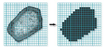
The images are then processed and a variety of morphological parameters are calculated.
Advanced graphing and data classification options in the Morphologi software ensure that extracting the relevant data from your measurement is straightforward, via an intuitive visual interface.
Individually stored grayscale images for each particle provide qualitative verification of the quantitative results.
Automated imaging for advanced particle characterization