Differential Scanning Fluorimetry (DSF)
Rapid insights into protein stability, formulation, and biomolecular interactions

Rapid insights into protein stability, formulation, and biomolecular interactions
Differential Scanning Fluorimetry (DSF) is a rapid biophysical technique used to assess protein conformational stability by tracking changes in fluorescence as a sample is subjected to controlled thermal stress. As the temperature increases, thermal energy disrupts the protein's native three-dimensional structure, exposing internal regions that were previously hidden within its hydrophobic core.
In traditional extrinsic DSF this structural unfolding is detected using external reporter dyes that will bind to newly exposed hydrophobic regions. Label free intrinsic DSF relies on the natural fluorescence of the aromatic amino acids such as tryptophan within the protein. As the sample unfolds the aromatic amino acid transitions from a buried non-polar environment to an exposed polar environment, this changes the natural fluorescence emission spectrum of the protein.
Intrinsic differential scanning fluorimetry relies on the amino acid tryptophan, which absorbs UV light at 280 nm and emits fluorescence between 300 nm and 400 nm.
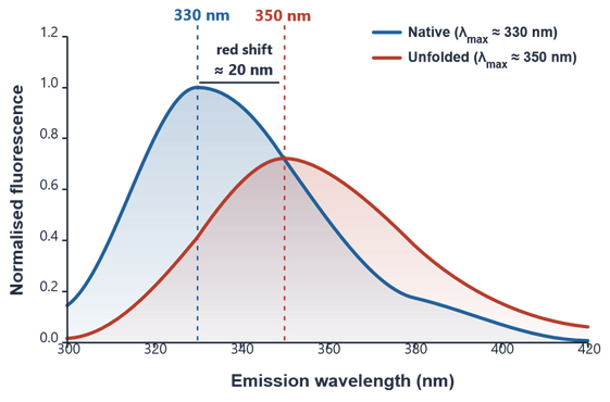
In a native, folded protein, tryptophan residues typically reside in the hydrophobic core and emit light peaking around 330 nm. As thermal stress forces the protein to unfold, these buried residues become exposed to the surrounding polar aqueous environment, causing the emission spectrum to shift toward 350 nm.
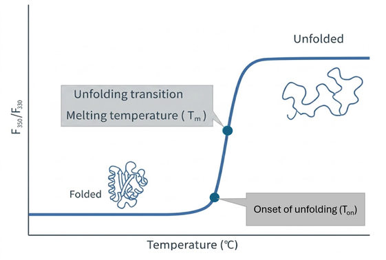
By recording the ratio of these two emission wavelengths (F350/F330) across a controlled temperature ramp, the system plots a sigmoidal thermal unfolding curve. Mathematical analysis of this curve identifies the onset of unfolding (Ton) and the midpoint melting temperature (Tm), where 50% of the protein population has unfolded.
Because a higher Tm reflects greater resistance to heat-induced denaturation, DSF allows researchers to quickly rank candidate molecules, screen buffer conditions, and evaluate candidate stability across drug development pipelines.
Furthermore, when combined with light scattering methods (such as static light scattering or dynamic light scattering), intrinsic DSF helps distinguish between conformational unfolding and physical aggregation, offering a comprehensive stability profile of a protein in solution.
Differential Scanning Fluorimetry (DSF) measures protein thermal stability by monitoring conformational changes as temperature increases. The difference between extrinsic and intrinsic DSF comes down to if a report dye is added or not.
| Feature | Extrinsic DSF | Intrinsic DSF |
|---|---|---|
| Mechanism | Uses an added fluorescent dye (e.g., SYPRO Orange) that binds to exposed hydrophobic regions during unfolding. | Label-free technique that tracks changes in native fluorescence from aromatic amino acids (Trp/Tyr). |
| Sample Prep | Requires dye addition and optimization. | Zero labelling; measures proteins in their native buffer. |
Limitations of Extrinsic DSF:
Limitations of Intrinsic DSF:


Next generation biophysical screening at scale